 I saw the funniest presentation ever at the SPIE Medical Imaging conference last week in San Diego. Few researchers from HP were showing a new method for annotating medical images. I wish they had talked to a radiologist before presenting the paper and making a fool of them selves. You have see it to understand what I mean. Here is the link to their video podcast for some example image annotations: http://www.hpl.hp.com/personal/I-Jong_Lin/hp_labs_experimental_podcast.xml
Now that you have seen the video, here is what want a radiologist to do:
- To annotate an image first the radiologist identifies an image with abnormality.
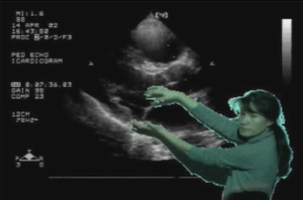
- Then he has to become a weather man and stand in front of a blue screen and a video camera.
- Now he has to capture video of himself pointing to the virtual abnormality on the image.
- Then some how save this video - which is not DICOM
- Then send to a clinician - again not using the RIS or HIS but some other way.
- Lastly get fired because you wasted whole day annotating a single image and rest of the work was delayed.
On average a radiologist spends 90 seconds reading a radiograph and about 7 minutes interpreting a CT study. What ever annotation methods engineers and programmers develop has to be part of the 90 seconds for a radiograph and 7 minutes for a CT examination.
Advice for Engineers and Software developers: Please! Please! Consult the users (radiologist) before designing a system.
No comments:
Post a Comment